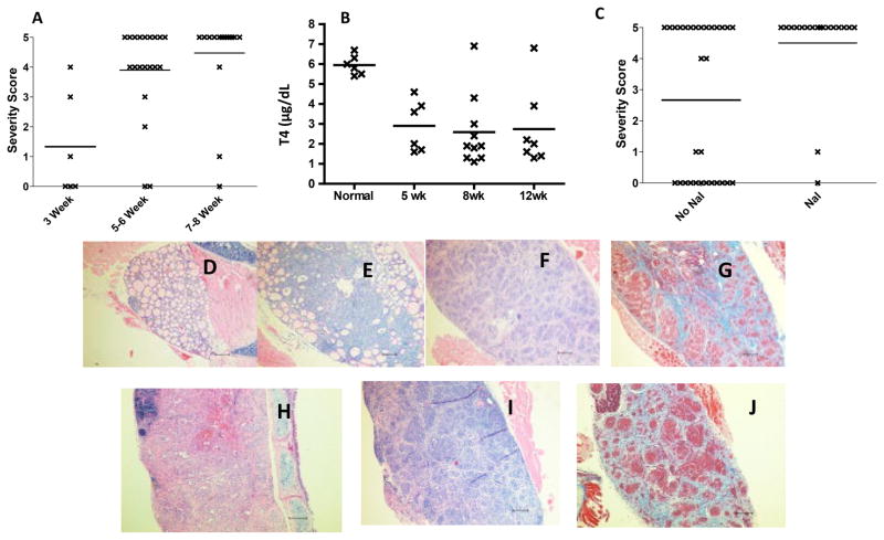
Figure 1.
Rapid development of severe TEC H/P in IFN-γ−/− CD28−/− NOD.H-2h4 mice. A) IFN-γ−/−CD28−/− mice (both sexes) were given NaI water at 7–8 wk of age. Thyroids were removed 3, 5–6 or 7–8 wk later as indicated. TEC H/P severity scores of individual mice are shown. N =6 (3 wk); 20 (5–6 wk) and 19 (7–8 wk). B) Serum T4 levels for some mice in Fig. 1A. N=6 (normal mouse); N= 6 (5 wk); N= 9 (8 wk) and N= 6 (12 wk). Serum T4 is significantly reduced in all groups compared to normal mice (p<0.05) C) IFN-γ−/− CD28−/− mice (both sexes) were maintained on plain water (no NaI) or given NaI water at 7–8 wk of age. Thyroids were removed when mice were 5–6 mo old. Mice given NaI water had a significantly higher incidence of severe (4–5+) TEC H/P compared to those not given NaI water (p< 0.01). N=30 (no NaI) and N =18 (NaI). D -J) H&E (D-F, H, I) or Trichrome (G, J) stained sections of thyroids from IFN-γ−/− CD28−/− NOD.H-2h4 mice given NaI water for 3 (D, 0+ severity), 5 (E, 4+ severity), or 8 wk (F,G 5+ severity) or from mice maintained 6 mo on plain water (H, I, J, 5+ severity). Note the extensive proliferation of thyrocytes with virtually complete obliteration of normal follicles, and fibrosis (blue). Pictures are representative of at least 6 mice examined in D, G and J and 20 or more for other panels. Magnification: X100.

