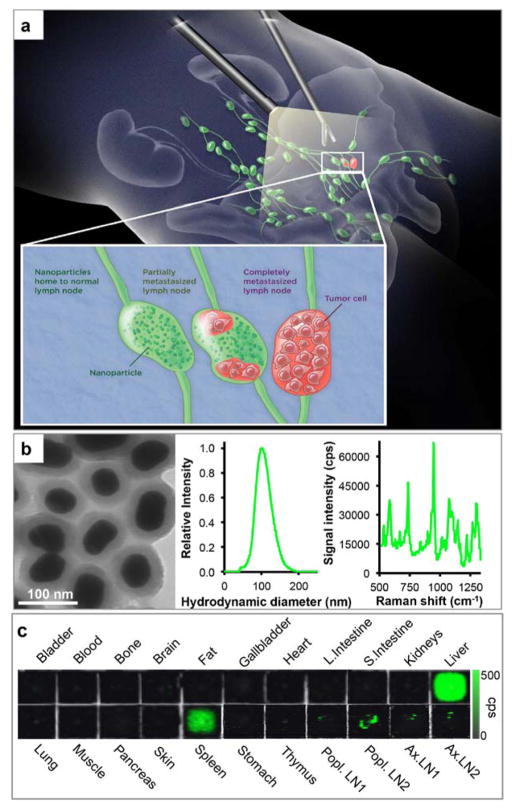
Figure 1. SERRS-based Raman imaging for the detection of prostate cancer lymph node metastases.
a. Concept. SERRS nanoparticles are injected intravenously, reach lymph nodes via interstitial-lymphatic fluid transport and internalize into macrophages within healthy lymph nodes. Magnified inset: In normal lymph nodes (left) the nanoparticles are taken up avidly by resident macrophages. If a lymph node is either partially (middle) or completely (right) invaded by metastatic prostate cancer cells, the resident macrophages are replaced by tumor tissue, resulting in a marked decrease in nanoparticle uptake. b. Transmission electron micrograph (left), nanoparticle size distribution determined with nanoparticle tracking analysis (middle), and Raman spectrum of the SERRS nanoparticles upon excitation with a 785-nm laser (right). c. Tissue distribution of the SERRS nanoparticles in a representative healthy mouse. Raman signal of the 950 cm−1 in counts per second.

