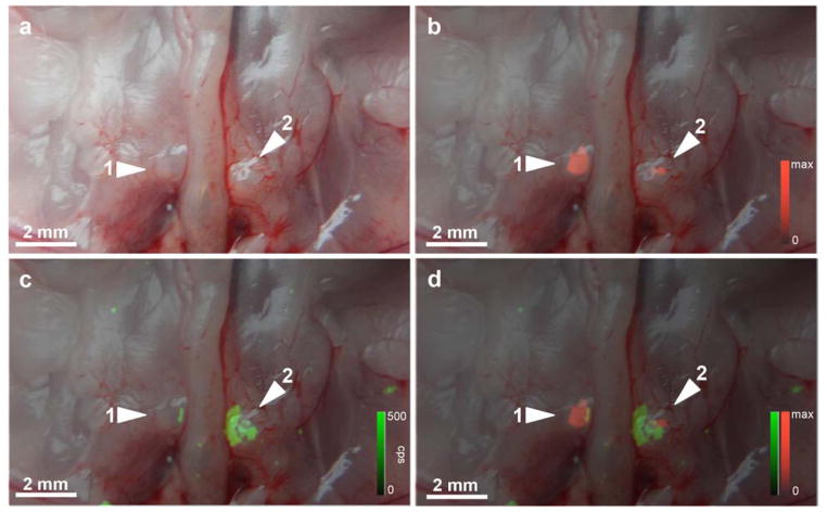
Figure 2. In situ fluorescence and Raman imaging of lymph node metastases in an orthotopic mCherry-transduced PC-3 prostate cancer mouse model.
Animals were injected 16–18 h prior i.v. with SERRS nanoparticles. a. Photograph of the retroperitoneum after radical prostatectomy. Medial iliac lymph nodes are indicated by arrowheads. b. Fluorescence/white-light overlay of the retroperitoneum shows the fluorescence signal of mCherry-transduced PC-3 cells (red pseudocolor) in the medial iliac lymph nodes, indicating the presence of lymph node metastases. c. Raman (green pseudocolor)/white-light overlay and d. Raman/mCherry-fluorescence/white-light overlay of the same lymph nodes shows SERRS signal only in those areas of the lymph nodes that are not replaced by tumor cells.

