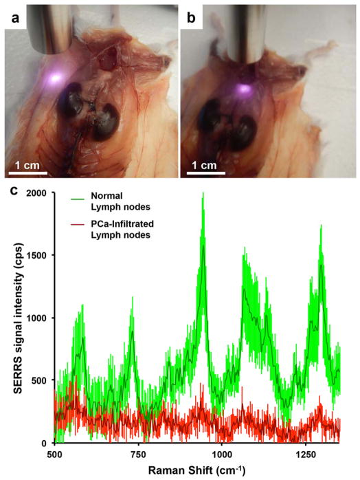
Figure 5. Feasibility of detecting lymph node metastases with a hand-held Raman spectroscopy scanner in near real-time.
In situ hand-held Raman scanning (785 nm, 10-mW laser power, 100-ms acquisition time) was performed on a. healthy subiliac lymph nodes and, b. metastasized medial iliac lymph nodes. c. While the Raman fingerprint of the SERRS nanoparticles is detected in the healthy lymph nodes (green spectrum), a significant (p < 0.05) decrease in SERRS signal of the 950 cm−1 peak was found in the diseased lymph nodes (red). Data represent at least four different experiments and are presented as mean normalized SERRS spectra±SD.

