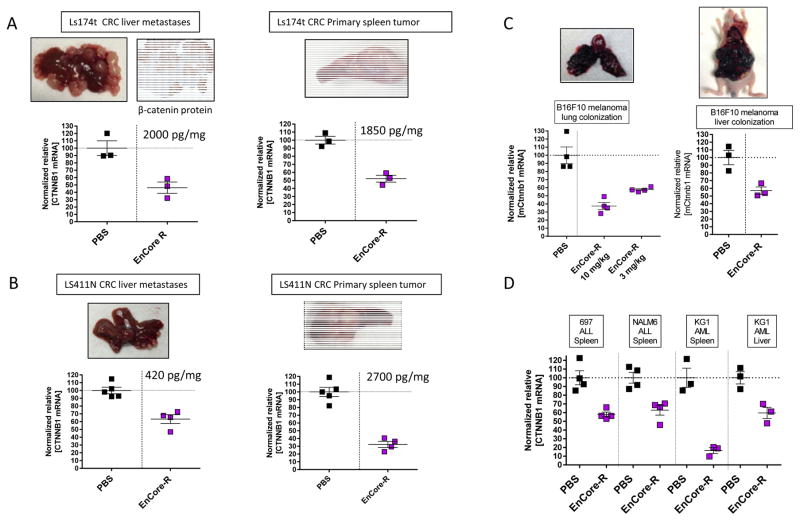
Figure 3. EnCore-R delivers CTNNB1 DsiRNA to metastatic and disseminated cell line-derived tumors.
Colorectal cancer (CRC) liver metastases were generated by surgically implanting Ls174t (A) and LS411N (B) cells in spleens of nude mice. The splenic primary tumor and liver metastases were independently evaluated for CTNNB1 mRNA 24 hours after dosing EnCoreR/CTNNB1 or PBS. Representative tissue images to show tumor morphology are included, as well as a representative 4× immunohistochemistry image for human β-catenin protein to confirm the identity of the LS174t tumor in (A). Also indicated on the plots are the mean [DsiRNA] levels measured in the primary spleen and metastatic liver tumors at the time of harvest. In (C), murine B16F10 melanoma cells were injected intravenously into wild-type mice and allowed to colonize the lung (left panel) or injected hydrodynamically to form large tumors in the liver (right panel). Mouse Ctnnb1 (mCtnnb1) was measured in the tumor 24 hours after administering EnCore-R/CTNNB1 or PBS. In (D), human acute lymphoblastic leukemia (ALL) or acute myelogenous leukemia (AML) cells were injected intravenously into immunocompromised mice. After allowing for the tumor cells to colonize the spleen or liver, as indicated, EnCoreR/CTNNB1 DsiRNA was administered and mRNA knockdown was evaluated after 24 hours. n=3 or 4 animals per cohort in all panels. All cohorts received a single dose of 10 mg/kg except where indicated; p<0.05 vs. PBS for all experiments shown in this figure.

