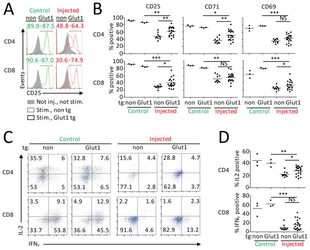
FIGURE 4. Reduced Glut1 expression contributes to dysfunction of T cells from leukemia-bearing hosts.
Splenocytes from not injected mice with T cells with (n=3) or without (n=3) Glut1 tg and FL5.12-injected mice with T cells with (n=20) or without (n=10) Glut1 tg were stimulated over-night with anti-CD3. (A) Representative data of CD25 expression. Numbers represent % of positive cells in no tg and Glut1 tg animals. (B) CD25, CD71 and CD69 expression measured with flow cytometry. (C,D) Production of IL-2 and IFNγ was assessed after additional 5h stimulation with PMA/Ionomycin. Numbers in each quadrant represent % of events. * P < 0.05, ** P < 0.01 and *** P < 0.001.

