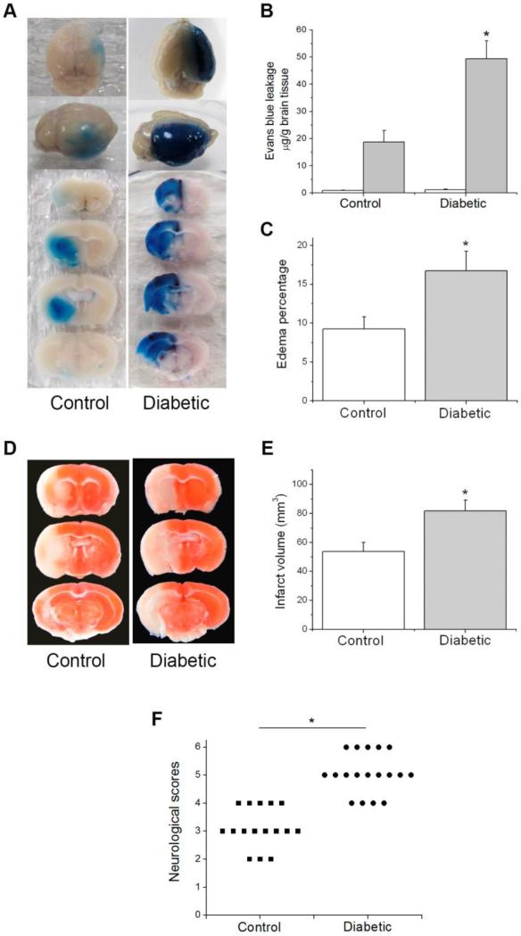
Fig. 1.
Effect of diabetes on ischemia/reperfusion-induced brain injury. Control and diabetic mice were subjected to 90 min MCAO followed by 24 h reperfusion. (A) Representative images of EB extravasation in a whole brain and coronal sections. (B) Quantification of EB leakage in contralateral and ipsilateral hemispheres (n=5). White bars, contralateral hemisphere; dark bars, ipsilateral hemisphere. (C) Quantification of brain edema percentage (n=6 (control), 8 (diabetic)). (D) Representative TTC staining images of brain sections. (E) Quantification of infarct volume estimated by TTC stained sections (n=6 (control), 8 (diabetic)). (F) Quantification of neurological deficit scores rate (n= 16 (control), 18 (diabetic)). Values are means ± SD, *p< 0.05 vs. control animals.

