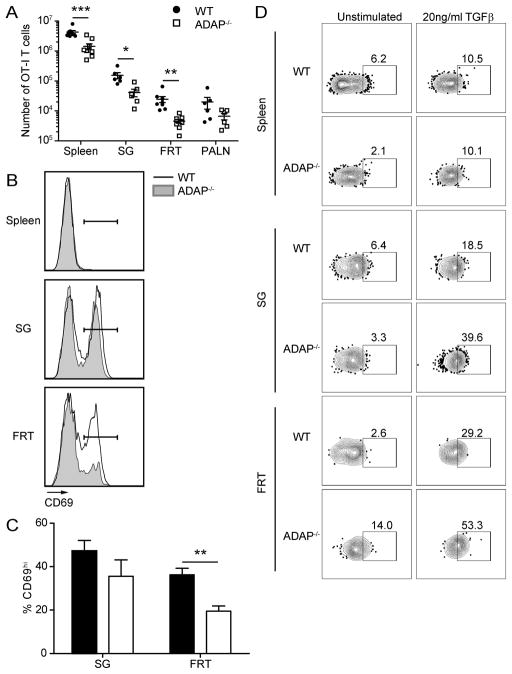
Figure 5. ADAP is required for optimal CD8 TRM cell presence at memory time points.
Naïve WT (CD45.1/2) and ADAP−/− (CD45.2) OT-I T cells were cotransferred into CD45.1 hosts and challenged with VSV-OVA. Tissues were harvested and single cell suspensions were stained for cell surface markers at D33+ challenge. (A) Number of WT (black circles) or ADAP−/− (open squares) OT-I T cells in the spleen, SG, FRT or para-aortic lymph node (PALN) after VSV-OVA challenge. (B) CD69 staining from WT (black line) and ADAP−/− (grey shaded) OT-I T cells from spleen, SG and FRT. Gate represents CD69hi population. (C) Percentage of wild-type or ADAP−/− OT-I cells with CD69hi staining. The results in (A) are compiled from 3 independent experiments, with at least 4 mice per experiment (± SEM). The results in (C) are compiled from 2 independent experiments, with at least 3 mice per experiment (± SEM). (D) Single cell suspensions from the indicated tissues were prepared from mice 140 days post VSV-OVA infection and treated with control media or media containing 10 ng/ml TGFβ for 20 min and then stained for phospho-SMAD2/3. **, p < 0.01.

