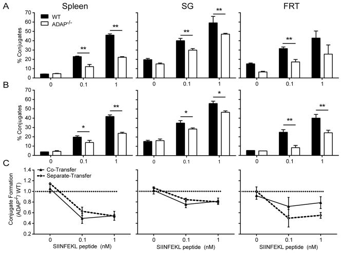
Figure 8. Impaired T:APC conjugate formation by ADAP−/− TRM cells isolated from NLTs.
Naïve WT (CD45.1 for single-transfer and Thy1.1 for co-transfer) or ADAP−/− (CD45.1) OT-I T cells were co-transferred (A) or separately transferred (B) into CD45.2 hosts, and challenged with VSV-OVA. On Day 120–160 following infection, the indicated tissues were isolated and processed to generate single cell suspensions that were mixed with DC-enriched splenocytes that were pre-pulsed with the indicated concentrations of SIINFEKL peptide. The formation of T:APC conjugates was determined by flow cytometry as described in Materials and Methods. (A) and (B) depict T:APC conjugate formation efficiency in a representative experiment from co-transferred (A) and separately transferred mice (B). Compiled results from 4 independent experiments, normalized to the conjugate efficiency of WT memory cells in each assay, are shown in (C).

