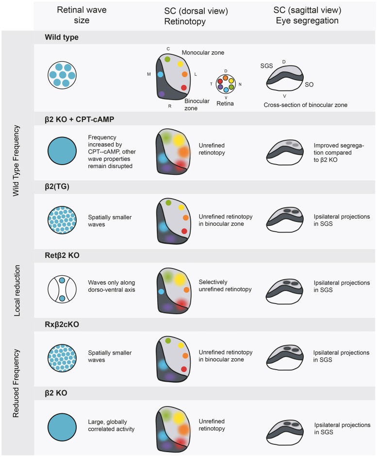
Figure 2.
Manipulations of spontaneous activity frequency and wave size and the consequences for retinotopy and eye-specific segregation in the SC. The SC has a binocular and a monocular region, and contains a retinotopic map in a mirror image of the retina. The most superficial layer of the SC is the stratum griseum superficial (SGS), which is targeted only by axons from the contralateral eye. The stratum opticum (SO) contains ipsilateral projections in wild type animals. Wild type: retinal activity in the wild type mouse. β2 knockout+ cAMP-CPT: this manipulation increases frequency to wild type levels (Burbridge et al., 2014), rescuing eye specific segregation but not retinotopy. β2 (TG): truncated waves as in the β2 (TG) mouse disturb eye-specific segregation (Xu et al., 2011). Retβ2-KO: partially disrupting wave activity also has spatially selective consequences for retinotopy (Burbridge et al., 2014). Rxβ2-KO: reducing wave frequency and size disturbs segregation (Xu et al., 2015) and retinotopy in the binocular zone. β2 KO: the whole body β2 knockout has low frequency activity over large areas of the retina, leading to unrefined retinotopy and disrupted eye specific segregation. SGS, stratum griseum superficiale; SO, stratum opticum; D, dorsal; V, ventral; C, caudal; R, rostral; T, temporal; N, nasal.

