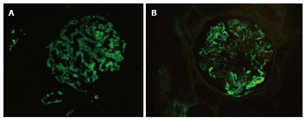
Figure 2.

Comparable staining pattern on immunofluorescence on frozen and paraffin embedded tissue. A case of dense deposit disease showing bright C3c deposition 3+ (0-3+ scale) on IF-F (A, FITC C3c, × 200). Note the comparative coarse granular capillary wall staining of C3c (3+) on paraffin embedded tissue section after enzymatic retrieval (B, FITC C3c, × 200). FITC: Fluorescein isothiocyanate; IF-F: Immunofluorescence on fresh frozen tissue.
