. 2016 Aug 30;57(6):1312–1323. doi: 10.3349/ymj.2016.57.6.1312

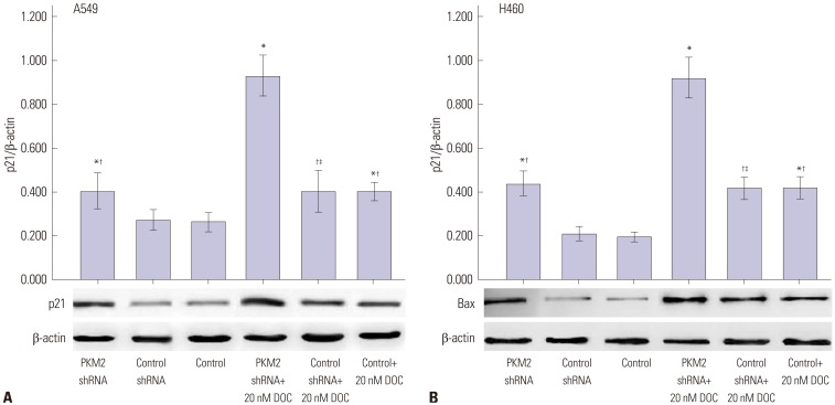
Fig. 5. p21 protein expression induced by 0 nM and 20 nM docetaxel in PKM2-shRNA transfected, shRNA-control transfected or non-transfected cells at 48 hrs. 48 hrs after transfection, all A549 (A) and H460 (B) cells, PKM2 shRNA-transfected (PKM2 shRNA), control-shRNA transfected (Control shRNA) or non-transfected (Control), were incubated with 20 nM docetaxel for up to 48 hrs, Western blot analyses were performed to detect the expression of p21. The graph depicts mean±SEM for three independent determinations of optical density of the p21 Western blot bands. *Compared to control cells with incubation of 0 nM docetaxel, p<0.05, Compared to PKM2 shRNA-transfected cells with incubation of 20 nM docetaxel, p<0.05, Compared to control shRNA-transfected cells with incubation of 0 nM docetaxel, p<0.05. PKM2, the M2 isoform of pyruvate kinase; DOC, docetaxel.

Fig. 5