
Figure 15.
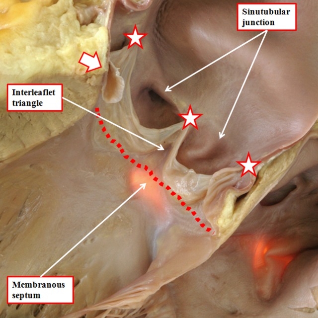
The outflow tract of the normal heart has been opened through an incision across the left coronary sinus of the aortic root. The membranous septum has been transilluminated from the right side. This shows how the triangular space between the right coronary and the nonadjacent sinuses of the root is filled by a fibrous wall. Similar triangles with fibrous walls are to be found beneath each of the valvar commissures, these being the points at which the leaflets join together at the sinotubular junction (white stars with dark borders). The relationships of these triangles are shown in Figure 16. Note how the virtual basal plane is created by joining together the basal attachments of the valvar leaflets (dotted line).