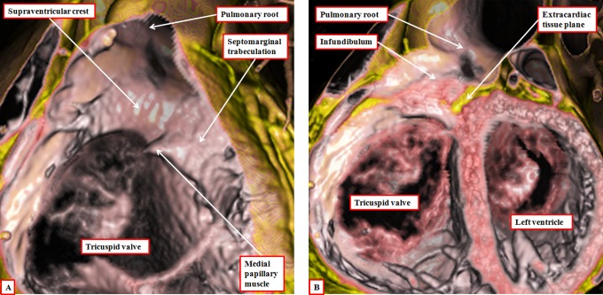
Figure 17.
The images show virtual dissections revealing the structure of the right ventricular infundibulum. The left panel (A) is made by cutting away the parietal wall of the right ventricle, showing how the posterior wall of the infundibulum is formed by the supraventricular crest. The larger part of this wall is no more than the inner heart curvature. The component adjacent to the aortic root, however, is the free-standing infundibular sleeve, the presence of which makes it possible to remove the root for use as an autograft in the Ross procedure. The right panel (B) shows this sleeve, revealing the tissue place that separates it from the aortic root.

