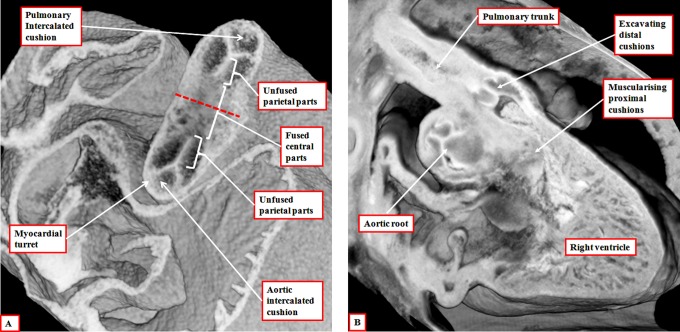
Figure 6.
The left panel (A) is prepared using an episcopic data set from a mouse embryo killed during the 13th day of development. The short axis of the intermediate part of the outflow tract is viewed from above, showing the developing primordiums of the aortic root. Note that, at this stage, the roots remained encased in a turret of outflow tract myocardium. The right panel (B) is from an episcopic data set prepared using a human embryo at Carnegie stage 20. The three parts of the outflow tract are shown, with the distal cushions excavating to form the leaflets of the pulmonary valve, and the surface of the fused proximal cushions muscularizing to form the subpulmonary infundibulum. At this stage, the intermediate and proximal parts of the outflow tract retain their myocardial walls.

