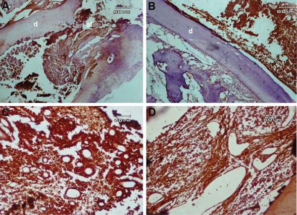
Fig.3.

Immunohistochemical staining with angiogenic factors. A. Immunostaining to evaluate the degree of angiogenesis in the root canal (original magnification: ×40), B. Expression of vascular endothelial growth factor (VEGF) in new vital tissue observed in the canal space (original magnification: ×40), C. High expression of VEGF in stromal and endothelial cells of the blood vessel walls. The intensity score was severe (I=4) (original magnification: ×100), and D. Expression of factor VIII in regenerated tissue observed in the canal space (I=3) (original magnification: ×100). d; Dentin.
