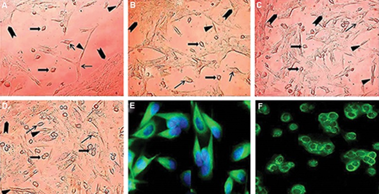
Fig.1.

Typical morphology of somatic and spermatogonial cells during the process of cultivation at A. 1, B. 2, C. 3, D. 4 days. Somatic cells including Sertoli (arrows), Leydig (arrowheads) and myoid (triangle) cells formed the feeder layer with grown spermatogonia at its surface (block arrow), E. Immunocytochemical evaluations of sheep testicular cells after 2-3 days culture using an antibody against vimentin, and F. Spermatogonia were identified by KIT immunocytochemical staining one week after culture initiation (scale bars A-D: 50 µm, and E, F: 40 µm).
