Fig. 1.

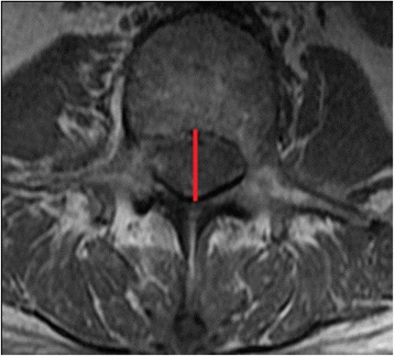
Measurement of midline anteroposterior (AP) spinal canal diameter at L1 to S1 using T1-weighted axial MRI scan (marked in red line) - at the cut where the entire bony canal ring could be seen and with the thickest pedicle width

Measurement of midline anteroposterior (AP) spinal canal diameter at L1 to S1 using T1-weighted axial MRI scan (marked in red line) - at the cut where the entire bony canal ring could be seen and with the thickest pedicle width