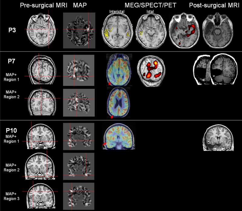
Fig. 4.
Unresected MAP+ region supported by noninvasive findings only (P3 and P7), or with unclear relevance (P10). P3 had one MAP+ region which was not resected. P7 had two MAP+ regions, one of which was resected. P10 had three MAP+ regions; only the right temporal one was included in the resection. P3 had both interictal and ictal MEG the yellow dipoles were modeled at the peak of interictal/ictal spikes. For P7, SISCOM was performed using z = 1.5

