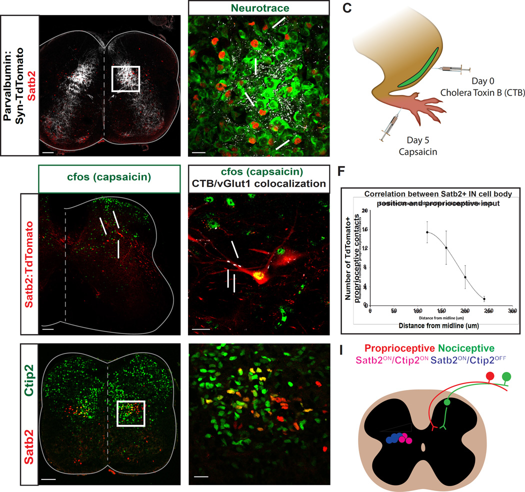
Figure 2. Satb2+ interneurons receive convergent sensory inputs and display heterogeneity along the mediolateral axis.
A, B. Proprioceptive fibers have a dense termination in the deep dorsal horn. Presynaptic terminals of proprioceptive fibers (white) were identified by crossing Parvalbumin-Cre to Rosa-LSL-Syp-TdTomato-deltaNeo (PV:Syn-TdTomato). Proprioceptive contacts (white) were identified on neurotrace+ cell bodies (green) of Satb2+ interneurons (red), highlighted by arrows. White box in A refers to high magnification image in B. P1 spinal cords in A, B. C. Schematic demonstrating experimental technique used to identify nociceptive relay neurons that also receive input from proprioceptive sensory fibers. Cholera Toxin-B was injected into the tibialis anterior (TA) muscle of Satb2:TdTomato animals to identify proprioceptive afferents. 5 days later, capsaicin was injected into the footpads and spinal cords were collected for labeling. D. c-Fos immunoreactivity (green) was used to identify cells in the spinal cord that are recruited following painful stimulation. Capsaicin was injected into the footpad of adult Satb2:TdTomato animals, as in C. Yellow cells that are co-positive for Satb2 (red) and c-Fos (green) are indicated by arrows. n=4 spinal cords. E. Satb2:TdTomato+ neurons (red) that receive multimodal sensory input were identified based on c-Fos immunoreactivity (green), and identification of proprioceptive synaptic terminals with vGlut1/CTB coexpression (white). F. Mediolateral position of Satb2+ interneurons corresponds to the number of contacts from proprioceptive fibers. Medial cells positioned closer to the midline had a greater number of contacts and lateral cells had fewer contacts. Quantified cells were binned by mediolateral position and the number of synaptic contacts is represented as mean +/− SEM. n=34 cells in 3 spinal cords. G, H. Ctip2 expression subdivides Satb2+ interneurons along the mediolateral axis. Medial Satb2+ interneurons (red) co-express Ctip2 (green), as indicated by yellow labeling in G, H. e15.5 spinal cords used for G, H. Scale bars in A, D, G, 100um. Scale bars in B, E, H, 20um. I. Summary diagram depicting features of Satb2+ interneurons. Left side: mediolateral organization of Satb2+ interneurons according to Ctip2 coexpression (blue/pink) and proprioceptive input (white wedge). Right side: multimodal sensory input from nociceptive (green) and proprioceptive pathways (red) onto Satb2+ interneurons (white).

