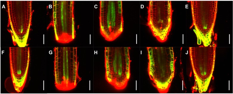
Figure 6.

Effects of electric field on cytokinin distribution during root tip regeneration. Temporal series of longitudinal median optical sections of representative root meristems expressing TCSn::GFP. Mock treatment (A)−(E) and aligned 2.5 V/cm electric field exposure (F)−(J) are shown, for both uncut roots (A, F) and individual regenerating roots at 0 (B, G), 1 (C, H), 2 (D, I), and 4 (E, J) days after treatment. green, GFP signal; red, propidium iodide counterstain; yellow, overlapping GFP and PI signals. Scale bars: 50 μm.
