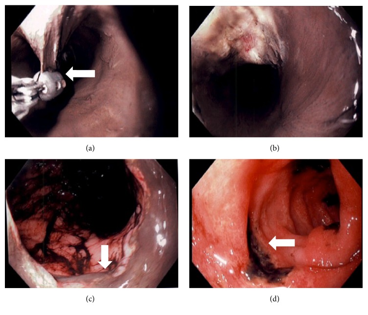
Figure 1.
Images demonstrating diffuse mucosal necrosis extending from the mid to distal esophagus in circumferential fashion. A biopsy forceps showing mucosal dissection (UL marked with arrow) with exposure the muscle layer (UR). Stomach demonstrating old digested blood throughout (LL) and discrete duodenal ulcers (LR marked with arrow) with ulcer base coated with blackish material suggesting ischemic changes.

