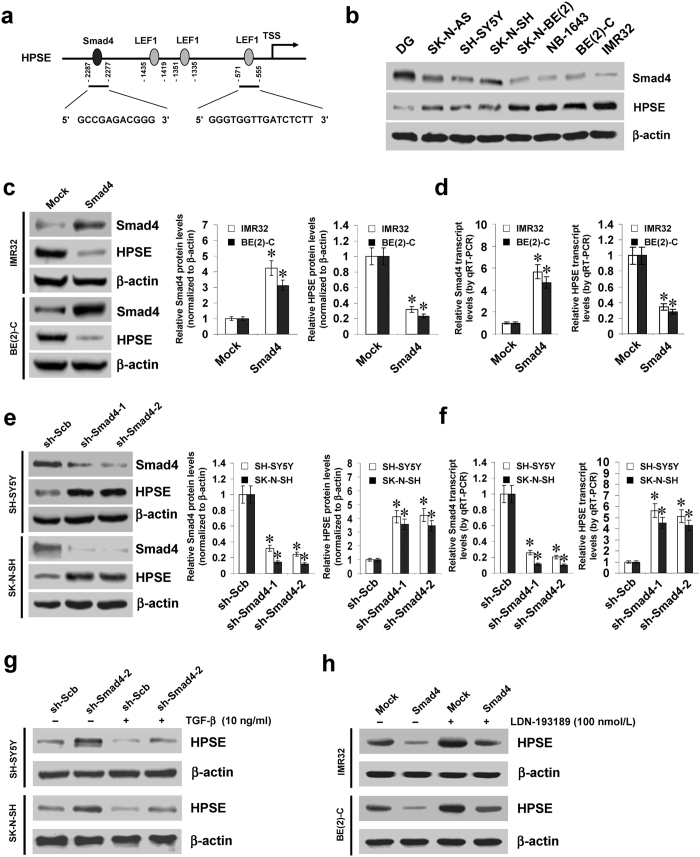
Figure 1. Smad4 represses the expression of HPSE in cultured NB cell lines.
(a) scheme of the potential binding sites of Smad4 and LEF1 within HPSE promoter, locating at bases −2287/−2277, −1435/−1419, −1351/−1335, and −571/−555 upstream the transcription start site (TSS). (b) western blot showing the expression levels of Smad4 and HPSE in normal dorsal ganglia (DG) and NB cell lines. (c,d) western blot and real-time quantitative RT-PCR indicating the protein and transcript levels of Smad4 and HPSE in IMR32 and BE(2)-C cells stably transfected with empty vector (mock) or Smad4. (e,f) western blot and real-time quantitative RT-PCR showing the protein and transcript levels of Smad4 and HPSE in SH-SY5Y and SK-N-SH cells stably transfected with scramble shRNA (sh-Scb) or shRNA specific for Smad4 (sh-Smad4). (g,h) western blot indicating the expression levels of HPSE in NB cells stably transfected with sh-Scb, sh-Smad4, mock or Smad4, and those treated with TGF-β or LDN-193189. *P < 0.01 vs. mock or sh-Scb.

