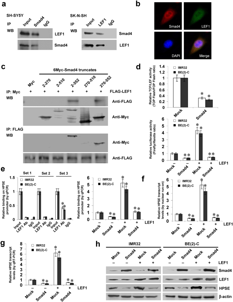
Figure 3. Smad4 represses the LEF1-facilitated transcription of HPSE in NB cells.
(a,b) Co-IP and immunofluorescence assays revealing the endogenous interaction between Smad4 and LEF1 in NB cells. (c) IP and western blot assays showing the interaction between Smad4 and LEF1 in IMR32 cells transfected with different Myc-tagged Smad4 truncates and FLAG-tagged LEF1 construct, running under the same experimental conditions (full-length blots are presented in Supplementary Figure S7). (d) dual-luciferase assay indicating the activity of TCF/LEF-responsive constructs TOP-FLASH and FOP-FLASH and the HPSE promoter reporter in NB cells stably transfected with empty vector (mock) or Smad4, and those co-transfected with LEF1. (e) ChIP and qPCR assay showing the enrichment of LEF1 on HPSE promoter in NB cells stably transfected with mock or Smad4, and those co-transfected with LEF1. (f,g) nuclear run-on and real-time quantitative RT-PCR assays indicating the HPSE transcript levels in NB cells stably transfected with mock or Smad4, and those co-transfected with LEF1. (h) western blot assay showing the expression of Smad4, LEF1, and HPSE in NB stably transfected with mock or Smad4, and those co-transfected with LEF1. *P < 0.01 vs. mock or IgG.

