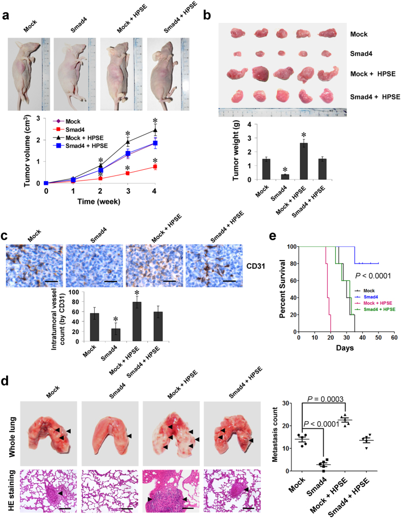
Figure 5. Smad4 suppresses the growth, metastasis and angiogenesis of NB cells in vivo.
(a) tumor growth curve of BE(2)-C (1 × 10⁶) stably transfected with empty vector (mock) or Smad4, and those co-transfected with HPSE in athymic nude mice (n = 5 for each group), after hypodermic injection for 4 weeks. (b) representation (top) and quantification (bottom) of xenograft tumors formed by hypodermic injection of BE(2)-C cells stably transfected with mock or Smad4, and those co-transfected with HPSE. (c) immunohistochemical staining (top) and quantification (bottom) of CD31 expression within tumors formed by hypodermic injection of BE(2)-C cells stably transfected with mock or Smad4, and those co-transfected with HPSE. Scale bars: 100 μm. (d) representation (left, arrowhead) and quantification (right) of lung metastasis in nude mice with injection of BE(2)-C cells (0.4 × 10⁶) stably transfected with mock or Smad4, and those co-transfected with HPSE via the tail vein (n = 5 for each group). Scale bars: 100 μm. (e) Kaplan–Meier survival plots of athymic nude mice with injection of BE(2)-C cells (0.4 × 10⁶) stably transfected with mock or Smad4, and those co-transfected with HPSE via the tail vein (n = 5 for each group). *P < 0.01 vs. mock.

