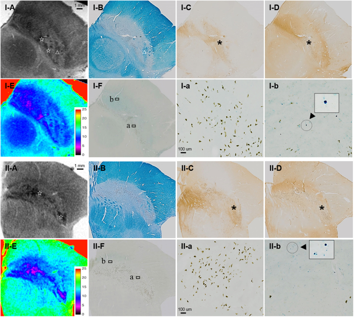
Figure 2. MRI and histology data from the middle of the substantia nigra (SN).
The clusters of neuromelanin (NM)-containing dopaminergic neurons were revealed as T2* hypointensity (☆) and low T2* values within the dorsolateral were revealed as hyperintense areas with a high tyrosine hydroxylase and low calbindin content (*). The regions of high NM content (a) were mostly distinct from those with a high iron content (b) as detected by Perls’ stain (Arrowhead: iron pigments). The clusters of NM exhibited better contrast in the older, 70-year-old female subject (II). Bundles of myelinated fibres in the ventrolateral SN also appear hypointense on T2*WI (Δ). A: T2*weighted imaging, B: Kluver-Barrera, C: Tyrosine hydroxylase immunohistochemistry, D: calbindin immunohistochemistry, E: T2* map (colour bar: T2* values), F: Perls’ Prussian blue.

