
Figure 6.
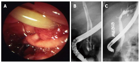
Duodenal ascariasis presenting as biliary colic. A: Duodenoscopy showing adult ascaride in the ampullary orifice; B: Endoscopic retrograde cholangiogram showing long linear filling defect in the common bile duct; C: Cholangiogram after extraction of worms from bile duct. Patient had immediate relief of biliary colic. Adapted from Khuroo[5].