
Figure 3.
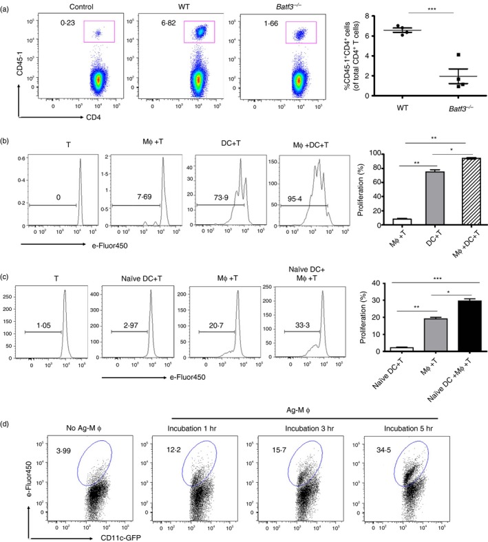
Splenic dendritic cells (DCs) acquired dead‐cell‐associated antigens from macrophages (Mφs) to amplify antigen‐specific CD4+ T‐cell proliferation. (a) Batf3 −/− or C57BL/6 mice were administered intravenously (i.v.) with 2 × 10⁶ CD62L+ Vβ5+ CD4+ CD45.1+ OT‐II T cells, followed by 4 × 10⁷ ovalbumin (OVA)/cells after 1 day. Three days later, the expansion of CD45.1+ CD4+ T cells was analysed by FACS. (b) C57BL/6 mice were i.v. injected with 2 × 10⁷ OVA/cells. One hour later, splenic Mφs and DCs were sorted and co‐cultured with 5 × 10⁵ eFluor‐450‐labelled CD4+ Vβ5+ Vα2+ OT‐II T cells individually or together. Three days later, the proliferation of CD45.1+ CD4+ OT‐II T cells was analysed with diluted eFluor‐450 dye. (c) Proliferation of co‐cultures of 1 × 10⁶ eFluor‐450‐labelled CD45.1+ OT‐II T cells with 2·5 × 105 DCs from naive mice (naive DCs), 2·5 × 105 Mφs from OVA/cells injected mice (Ag‐Mφs) or a combination of naive DCs and Ag‐Mφs. (d) Ag‐Mφs or no‐Ag‐Mφs were incubated with naive DCs. The kinetics of GFP + eFlour‐450+ DCs were determined by FACS. Data are shown as the mean ± SD of three to five mice in each group and are representative of three independent experiments. Two‐tailed Student's t‐test was used for data analysis. *P < 0·05, **P < 0·01, ***P < 0·001.