Figure 1.

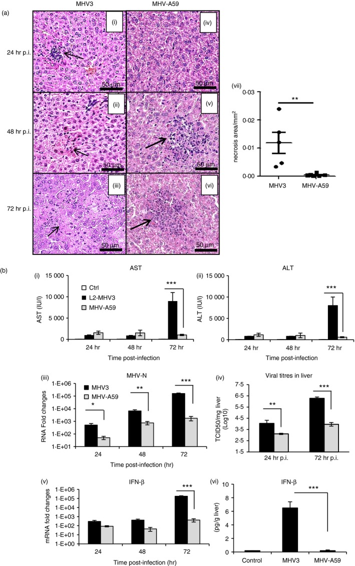
Mortality, hepatic damages and viral replication during murine hepatitis virus 3 (MHV3) and MHV‐A59‐induced hepatitis in C57BL/6 mice. Groups of six or seven C57BL/6 mice were intraperitoneally (i.p.) infected with 1000 TCID50 of MHV3 or MHV‐A59. Percentages of surviving mice were recorded at various times post‐infection (p.i.). (a) (i–vi) Histopathological analysis was conducted on livers at 24, 48 and 72 hr p.i. (vii) Histological analysis of inflammatory foci in liver from MHV3‐ or MHV‐A59 infected C57BL/6 mice. (b) Serum samples from infected mice were assayed for aspartate transaminase (AST) and alanine transaminase (ALT) activity from 24 to 72 hr p.i. (i, ii). MHV3 or MHV‐A59 replication in livers of infected mice was determined by analysis of the nucleoprotein (MHV‐N) RNA expression from 24 to 72 hr p.i. by quantitative RT‐PCR, and values represent fold change in gene expression relative to mock‐infected mice after normalization with HPRT expression (iii). Viral titration (TCID50) in liver was assayed at 24 and 72 hr p.i. (iv). mRNA fold increases for interferon‐β (IFN‐β) were evaluated by quantitative RT‐PCR in livers of MHV3‐ or MHV‐A59 infected mice. Values represent fold change in gene expression relative to mock‐infected mice (arbitrary value of 1) after normalization with HPRT expression (v). Production levels of IFN‐β were quantified by ELISA test at 72 hr p.i. in livers (vi). Results are representative of two different experiments. (*P < 0·05; **P < 0·01; ***P < 0·001).
