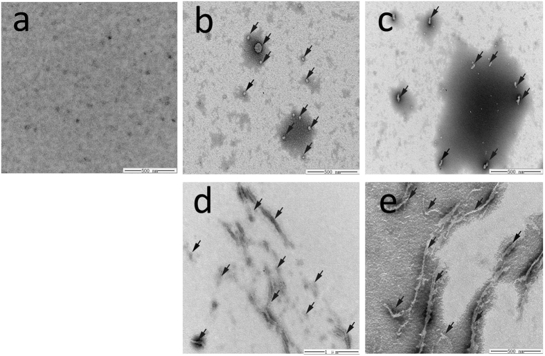
Figure 5. Morphology of KCTD1 aggregates reveals by transmission electron microscopy.
KCTD1 was incubated for 2 hours at 37 °C with or without 20 μM Cu2+. (a) Freshly purified KCTD1 presented no aggregates. (b,c) KCTD1 incubated without Cu2+ formed aggregates with irregular morphologies. (d,e) KCTD1 incubated with Cu2+ formed fibrillar aggregates. Irregular aggregates and fibrillar aggregation in (b–e) were indicated with black arrow mark.

