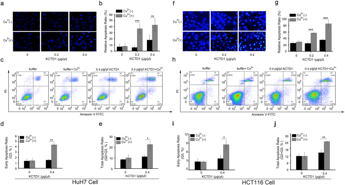
Figure 8. KCTD1 aggregates formed in the presence of Cu2+ induces more apoptosis than those formed in the absence of Cu2+ in HuH7 and HCT116 cell.
(a,f) Apoptosis was determined by observing chromatin condensation and fragment staining using Hoechst 33258 DNA staining. (b,g) Apoptotic nuclei were counted as a percentage of total nuclei in Hoechst 33258 DNA staining. (c,h) Typical images evaluated by flow cytometry. (d,i) The early apoptosis ratio and (e,j) total apotosis ratio (early apoptosis and late apoptosis) was quantified by Flow J software. The data shown are the mean ± s.d. of 3 independent experiments. *P < 0.05; **P < 0.01; ***P < 0.001; t-test.

