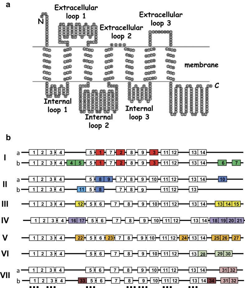
Figure 3. Motif organization of legume MLOs.
The figure shows the predicted topology of a typical MLO protein (a) and the schematic organization of the common and specific motifs for each MLO clade (b). Common and clade-specific motifs are represented by white and colored boxes, respectively. These motifs were identified by scanning the MLO sequences with the MEME suite software30 (Supplemental Table S1). Common and clade-specific amino acid motifs are listed in Tables 3 and 4 respectively. Localization of transmembrane domains is shown as dashed horizontal lines.

