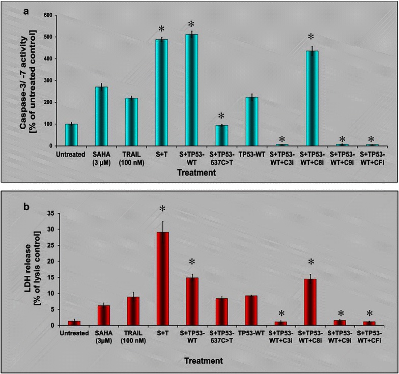
Fig. 4.

Apoptosis executioner caspase re-activation and LDH release in TP53-rescued ESS-1 cells. a Caspase-3 and -7 activation (Caspase-Glo 3/7 Assay) of TP53-WT- or TP53-637C>T-transfected ESS-1 cells that were treated with 3 µM SAHA. Different individual caspase inhibitors (C3i, C8i, C9i, CFi) were additionally applied together with SAHA to TP53-WT reconstituted cells as controls. Untreated, SAHA-, (S; 3 µM) and/or TRAIL-treated (T; 100 nM), and untreated TP53-WT-transfected cells served as further controls. b Measurements of LDH release by the CytoTox-ONE homogeneous membrane integrity assay of the samples analyzed in A for determination of the amount of cell cytotoxicity. Statistically significant differences compared to the SAHA-treated control were indicated in a and b
