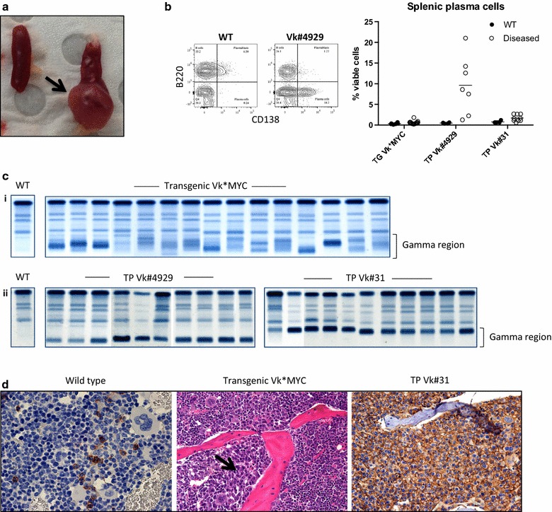
Fig. 1.

Disease characteristics of the transgenic and transplant Vk*MYC models. a Dissected spleen (left) wild type control (right) diseased Vk#4929 with a large plasmacytoma (arrow). b Plasma cells in the spleen of wild type and diseased Vk*MYC mice. Representative FACS plot is shown to the left: plasma cells are defined as CD138+B220− events and expressed as % viable cells. c Serum protein electrophoresis in individual mice at time of cull: (i) Transgenic Vk*MYC showing predominantly oligoclonal banding in the gamma region. (ii) Transplant Vk#4929 and Vk#31 showing monoclonal banding in the fast or slow gamma regions respectively. d BM trephine immunohistochemistry. Left CD138 staining showing normal numbers of plasma cells in wild type control. Middle H&E showing patchy plasma cell infiltrate in transgenic Vk*MYC (arrow). Right CD138 staining showing a diffuse infiltrate of plasma cells in transplant Vk#31
