
Fig. 6.
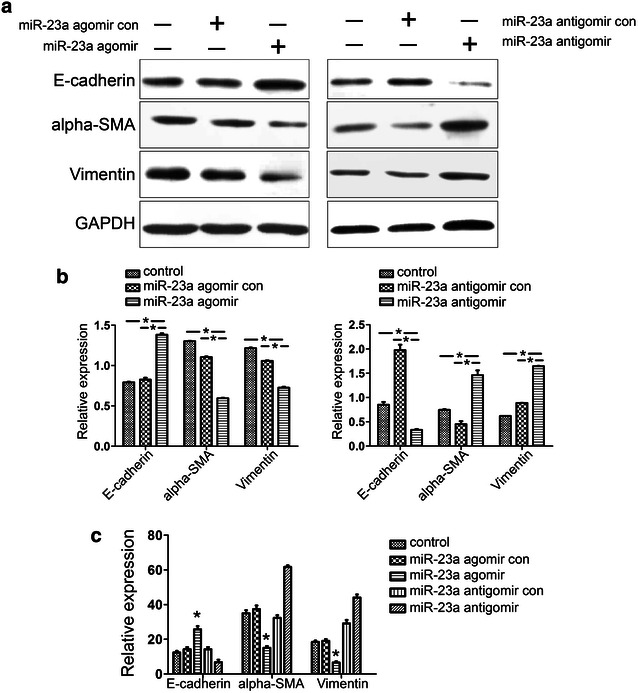
Expression of EMT markers when miR-23a is overexpressed in HEC-1-A cells. This experiment was conducted using treatment with 10 ng/mL TGFβ1 for 24 h, and the cells were then treated with the miR-23a agomir or antagomir investigate the effect of miR-23a on the EEC EMT. Non-treatment of TGFβ1 was used as the control group, and each group contained three samples. a, b Protein expression levels of the EMT markers. E-cadherin protein expression was increased when the HEC-1-A cells were treated with the miR-23a agomir but decreased when the cells were treated with the miR-23a antagomir. In contrast, the expression of both α-SMA and vimentin was decreased when the HEC-1-A cells were treated with the miR-23a agomir but increased when the cells were treated with the miR-23a antagomir. Top the gels (a); bottom the normalization graphs (b). c Up-regulation of miR-23a repressed the mRNA expression of EMT markers. The expression of mRNAs encoding α-SMA and vimentin was reduced in HEC-1-A cells treated with the miR-23a agomir but increased in cells treated with the miR-23a antagomir. However, the expression of E-cadherin mRNA was increased by the miR-23a agomir and decreased by the antagomir (n = 6 per group) (*P < 0.05)