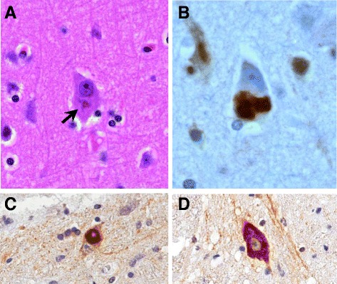
Fig. 2.

Neuronal and glial cytoplasmic inclusions immunoreactive for FUS define the pathology of both ALS-FUS and FTLD-FUS. Basophilic inclusions are present in neurons in ALS-FUS (arrowed) and can be viewed using H&E stain, X400 (a). Discrete neuronal inclusion immunoreactive for FUS associated with the P525L mutation, X400 (b). ALS-FUS inclusions in the anterior horn of spinal cord, both X40 Obj (c, d). Well defined, compact inclusions (c) or intense diffuse cytoplasmic staining (d) are commonly seen, often with nuclear clearance
