Figure 3:

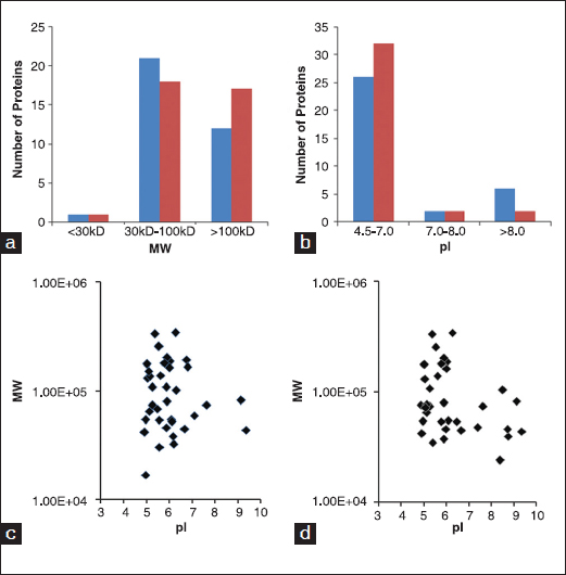
Molecular weight (MW) and isoelectric point (pI) distribution of extracellular matrix (ECM) proteins secreted by normal skin fibroblasts (NSFs) and hypertrophic scar fibroblasts (HSFs). (a) ECM proteins have been grouped into different MW bins according to their theoretical size. (b) ECM proteins have been grouped into different pI bins according to their theoretical value. Red bar stands for NSF and blue bar stands for HSF. (c and d) MW distributions of ECM proteins from NSFs and HSFS were plotted against their theoretical pIs, respectively.
