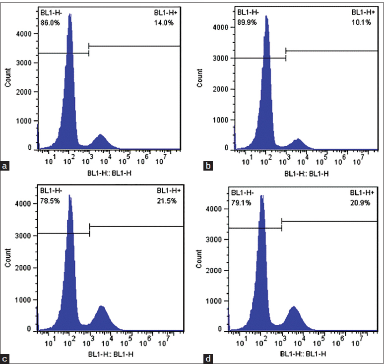
Figure 4:

Fluorescence activated cell sorting analysis of single-cell suspensions from wound tissue to measure the percentage of green fluorescence protein (GFP)-positive cells in the biological membrane-based, novel, excisional, wound-splinting model compared with the conventional model in C57BL/6 mice. (a and b) The percentage of GFP-positive cells was 14.0% on day 3 and 10.1% on day 5 in the conventional model. (c and d) The percentage of GFP-positive cells was 21.5% on day 3 and 20.9% on day 5 in the biological membrane-based, novel, excisional, wound-splinting model.
