
Fig. 2.
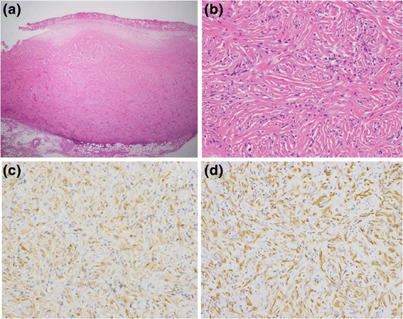
Pathological findings of a VATS-resected specimen (Case 3). Low-power image shows focal proliferation of cuboidal atypical cells with round nuclei and prominent nucleoli surrounded by fibrous tissue. Keratinization, plasmodesmata, and glandular construction were absent (a). High-power image of DMM (b). IHC demonstrated positivity for calretinin (c) and CAM5.2 (d)