
Fig. 1.
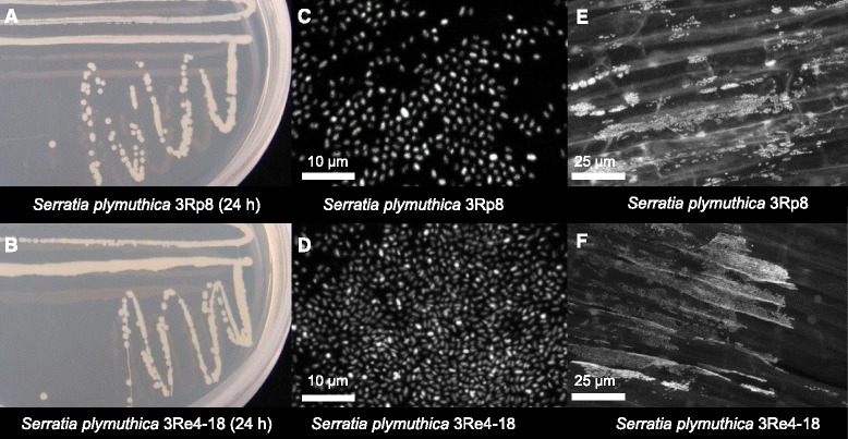
S. plymuthica 3Rp8 and 3Re4-18 on solid media and Confocal Laser Scanning Microscopy micrographs. a-b S. plymuthica 3Rp8 and 3Re4-18 grown on LB solid media after 24 h at 30 °C. Confocal Laser Scanning Microscopy micrographs: c and d show the cell morphology of pure cultures of 3Rp8 and 3Re4-18 after SYTO 9 green-fluorescent staining. e-f Fluorescence in situ hybridized 3Rp8 and 3Re4-18 colonizing the roots of young lettuce seedlings 1 week after inoculation in a gnotobiotic plant growth approach