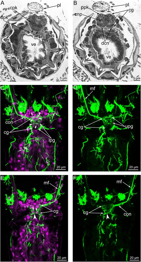
Fig. 7.

Organization of the central zone of the cerebral ganglion in Amathia gracilis. Semithin cross sections (a–b): the anal side is at the top, and the oral side is at the bottom. Anal view of Z-projections (c–f) of the lophophore after mono- and double staining for tyrosinated α-tubulin (green) and DAPI (magenta). a Cross section at the level of the tentacle bases. b Cross section at the level of the vestibulum. c Paired perikarya (double arrowheads) in the central zone of the cerebral ganglion. d Neurites of the central zone of the cerebral ganglion. e Paired perikarya (double arrowheads) and the chiasm (arrowhead) in the lower portion of the central zone. f Neurites forming a chiasm (arrowhead) in the central zone. Abbreviations: cg - cerebral ganglion; cnp - neuropil of central zone; cpk - perikarya of central zone; con - circum-oral nerve ring; dcn - cross neuropiles (commissures) in the distal zone; dpk - perikarya of distal zone; lpg - lower portion of the cerebral ganglion; mf - medio-frontal nerve of tentacle; mv - muscles of vestibulum; pl - pylorus; ppk - perikarya of proximal zone; tb - tentacle base; ve - vestibulum; upg - upper portion of the cerebral ganglion
