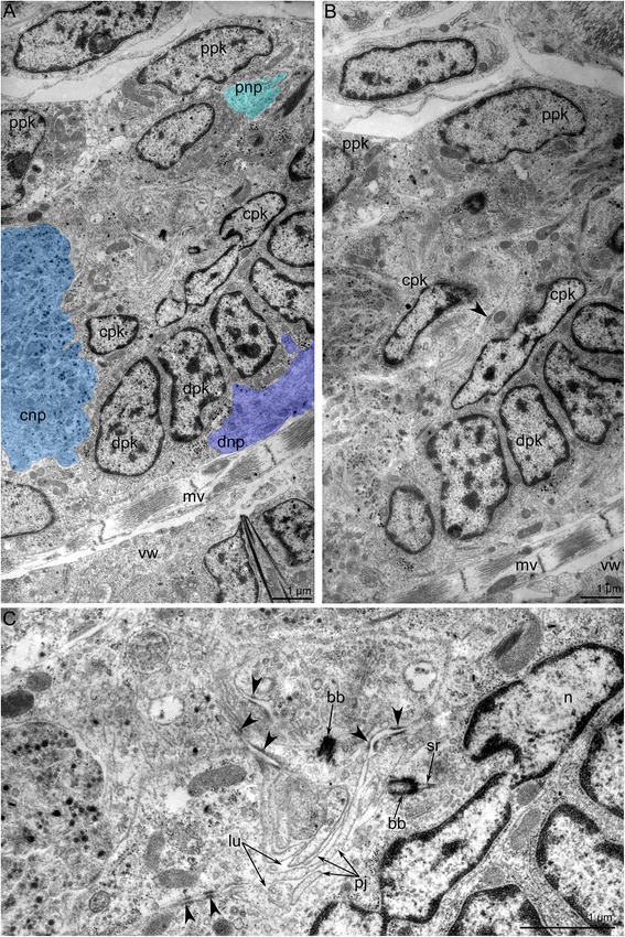
Fig. 9.

Ultrastructure of the cerebral ganglion in Amathia gracilis. Ultrathin cross sections; the anal side is toward the top, and the oral side is toward the bottom. a Three neuropiles of the cerebral ganglion marked by colors: proximal - cyan, central - blue, distal - violet. b Overview of all zones of the cerebral ganglion: perikarya of proximal, central, and distal zones. c Portion of the central zone - neuroepithelium. The lumen of the cerebral ganglion is filled with apical projections of the nerve cells, which connect via desmosomes (arrowheads) and bear the basal body. Abbreviations: bb - basal body; cnp - central neuropil; cpk - perikarya of central zone; dnp - distal neuropil; dpk - perikarya of distal zone; lu - lumen of cerebral ganglion; mv - muscle of vestibulum; n - nucleus; sr - striated rootlet; pj - apical projections; pnp - proximal neuropil; ppk - perikarya of proximal zone; vw - wall of vestibulum
