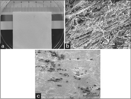
Figure 1:

Collagen scaffold morphology. (a) Macroscopic view of the collagen scaffold; (b) Scanning electron microscopy (SEM) image of the collagen scaffold. Scale bar =100 µm; (c) SEM image of the collagen scaffold conjugated with E7 peptide. Scale bar =100 µm.
