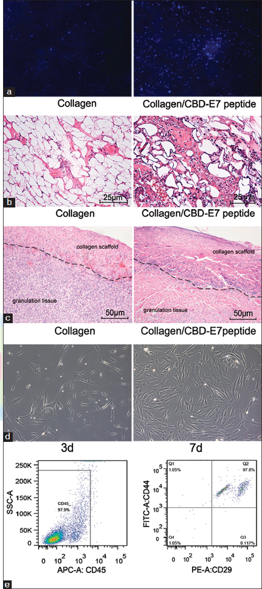
Figure 2:

Cellularization of collagen scaffolds. (a) Hoechst 33342 staining of the cells retained on the collagen with different treatments; (b) Cellular distribution in the deep fascia. The number of the cells infiltrated in the deep fascia in the collagen-binding domain (CBD)/peptide group was significantly higher than that in the Scaffold group (per field); (c) Histological evaluations of specimen tissues implanted with collagen scaffolds (left) and collagen/CBD-E7 peptide at day 7 post-surgery. The black curve shows the border between the material and the granulation tissue. (hematoxylin and eosin stain, ×200). (d) Cells derived from CBD-E7 Collagen scaffold at day 3 exhibit characteristic phenotypes of Mesenchymal stem cells (MSCs) (fibroblast-like growth and clumped together in a “swirl” pattern). (e) Fluorescence activated cell sorting (FACS) analysis of the cells remaining on the collagen scaffold.
