
Figure 1.
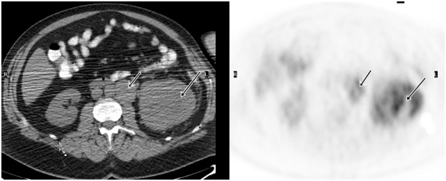
Demonstration of primary RCC and tumor thrombosis on FDG PET/CT. A 53-year-old man had a large left renal mass seen on the CT. FDG PET/CT showed increased, heterogeneous uptake of the mass in the left kidney. There was also tumor thrombosis in the renal vein, evidenced by FDG avid intraluminal lesion.