Fig. 1.
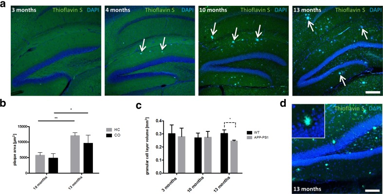
Development of amyloid-beta plaques in the hippocampus of APP-PS1 mice at different time points of AD progression. a Amyloid-beta plaques are first observed in the hippocampus of APP-PS1 mice at an age of 4 months and increased in number over time. b The area of Abeta antibody immunoreactive plaques significantly increased in the hippocampus of 13-month-old compared to 10-month-old AD animals. The same increase in amyloid-beta plaque load was observed in the cortex. c Analysis of the GCL volume in the dorsal dentate gyrus of the hippocampus revealed significantly decreased GCL volume specifically in 13-month-old APP-PS1 mice compared to WT (unpaired Student’s t test, p = 0,0213). d With progression of Abeta pathology, amyloid-beta plaques appear in the GCL of the dentate gyrus perforating the healthy tissue structure. Arrows show amyloid-beta plaques stained with Thioflavin S. DAPI was used to stain cell nuclei. Two-way ANOVA with Tukey’s multiple comparisons test (b) and unpaired Student’s t test (c) was performed (n = 3/group). Scale 200 μm (a), 100 μm (d)

