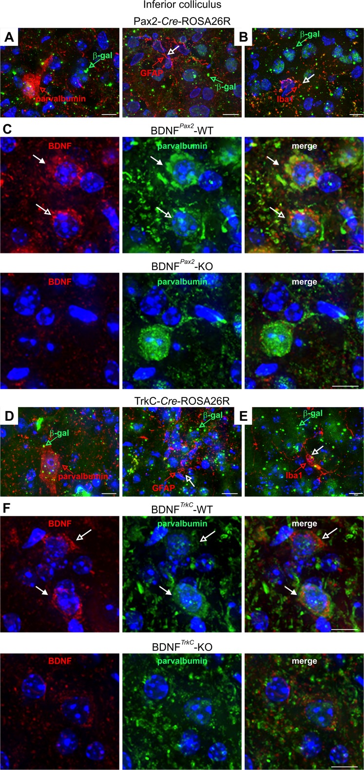
Fig. 2.
Immunohistochemistry of the IC of Pax2-Cre-ROSA26R and TrkC-Cre-ROSA26R mice and BDNFPax2 and BDNFTrkC WT and KO mice. a, d Immunostaining with anti-β-galactosidase (β-gal, green) and anti-parvalbumin (red) showing coexpression of β-galactosidase and parvalbumin in IC sections from both Pax2- and TrkC-Cre-ROSA26R mice. b, e Co-immunostaining with anti-β-galactosidase (β-gal, green) and either the oligodendrocyte marker anti-GFAP (red, open arrow) or the microglia marker anti-Iba1 (red, open arrow) in IC sections from both Pax2- and TrkC-Cre-ROSA26R mice shows no coexpression of β-galactosidase and GFAP or Iba1. Therefore, β-galactosidase is detected in neuronal cells. c, f Immunohistochemistry of IC sections stained with anti-BDNF (red) and anti-parvalbumin (green) antibodies, showing BDNF immunoreactivity in PV-positive and PV-negative neurons in BDNFPax2-WT (c, upper row) and BDNFTrkC-WT (f, upper row) mice. Closed arrows indicate cells positive for BDNF (red) and parvalbumin (green). Open arrows indicate cells expressing only BDNF (red). The specificity of the BDNF antibody is shown by the lack of BDNF immunostaining in BDNFPax2-KO and BDNFTrkC-KO mice (c, f, lower rows). Scale bars = 10 μm

