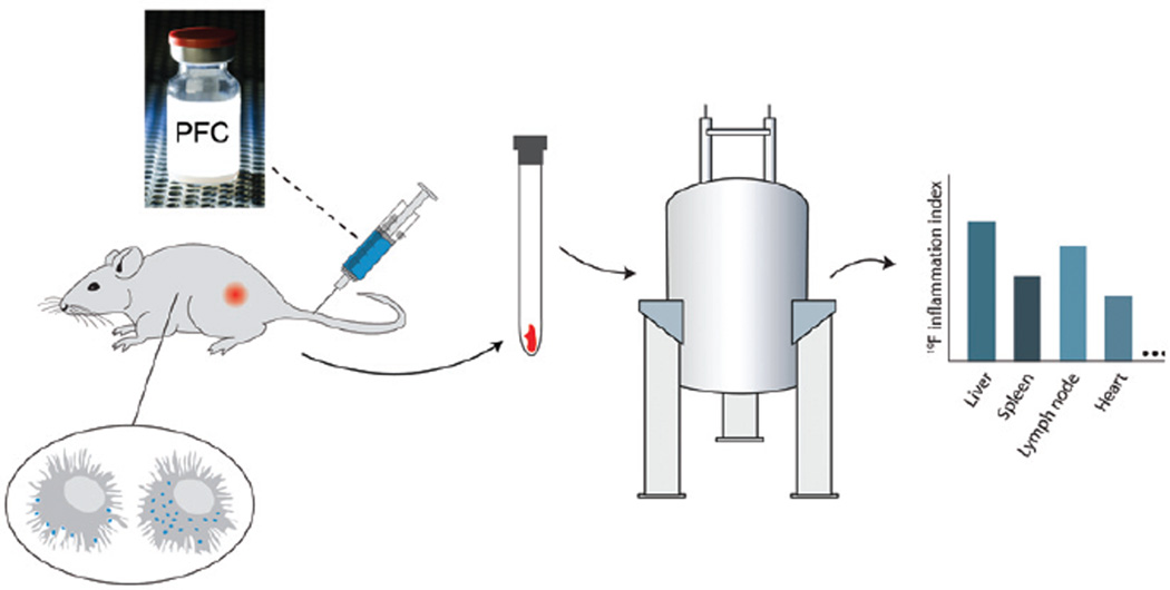
Figure 1. Overview of inflammation quantification of intact tissue samples using 19F NMR.
PFC emulsion is injected i.v. and is taken up by monocytes and macrophages. These labeled cells participate in inflammatory events in vivo resulting in an accumulation of 19F at inflammatory loci. Conventional 19F NMR spectroscopy of panels of intact tissue samples is used to measure histogram profiles of the inflammatory index (IFI), proportional to tissue macrophage burden.

