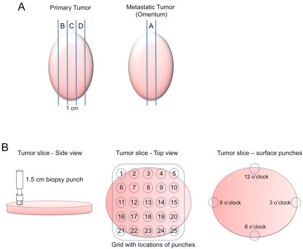
Figure 1. Schematic of sample collection.
(A) After debulking, a one centimeter slice was taken from the center of the primary and metastatic tumor along the longest diameter. These middle slices were designated Slice A from the metastasis and Slice C from the primary tumor. Additional 1-cm slices from either side of the middle slice were taken when possible from the primary tumor (slices B and D). (B) These slices were aligned with a grid allowing for up to 25 biopsy punches to be taken from each slice with a 2-cm spacing from the center of one punch to the center of the adjacent punch. Additionally, four punches were taken around the periphery of the tumor slice at 12, 3, 6 and 9 o'clock (punches 26, 27, 28 and 29). A peripheral blood sample was also collected from which mononuclear cells were isolated.

