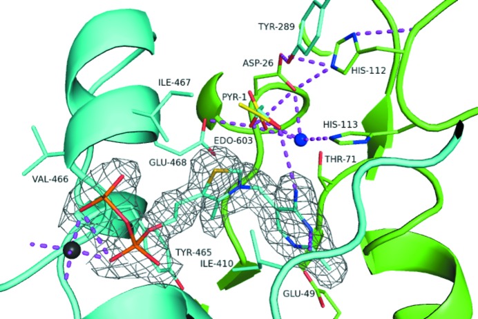
Figure 3.
Cartoon and stick depiction of the active site. Residues of one monomer are coloured cyan; residues of the other monomer are coloured green. The magnesium ion (dark grey) and water molecules (blue) are represented as spheres. The 1,2-ethanediol (EDO) and TPP of the cyan chain are shown as stick models and are coloured by atom. Pyruvate (PYR, yellow) has been overlaid following superposition of Z. mobilis PDC (PDB entry 2wva, chain F) and lies on top of the EDO. The 2F o − F c density (grey) surrounding the TPP is shown contoured at 1σ.

